

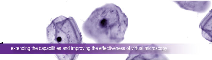
Virtual Microscopy can be defined as an artificial microscope environment, created on a personal computer, that when presented to a user has the “look-and-feel” of a real microscope.
A Virtual Microscopy system consists of two main parts: a scanning system, which digitises the microscopic specimens, and a client, which is used to browse and examine the digital specimens. The Virtual Microscopy project is broadly engaged with a number of enabling technologies and application areas capable of enhancing current best-practice in laboratory based pathology. Automated specimen digitisation and analysis is performed using a high-magnification microscope with an automated stage, together with a video camera that captures high-resolution images of the tissue and cells. These digital specimens can then be quickly and efficiently analysed on a high performance personal computer, or browsed remotely from a client computer.
This work has the potential to revolutionise a number of pathology applications including: high volume tests required for the early detection of cervical, lung, bladder and oral cancers; tele-pathology in remote areas; pathology and anatomy training; and laboratory quality assurance programs.